
 Dra. Lisbania Peguero

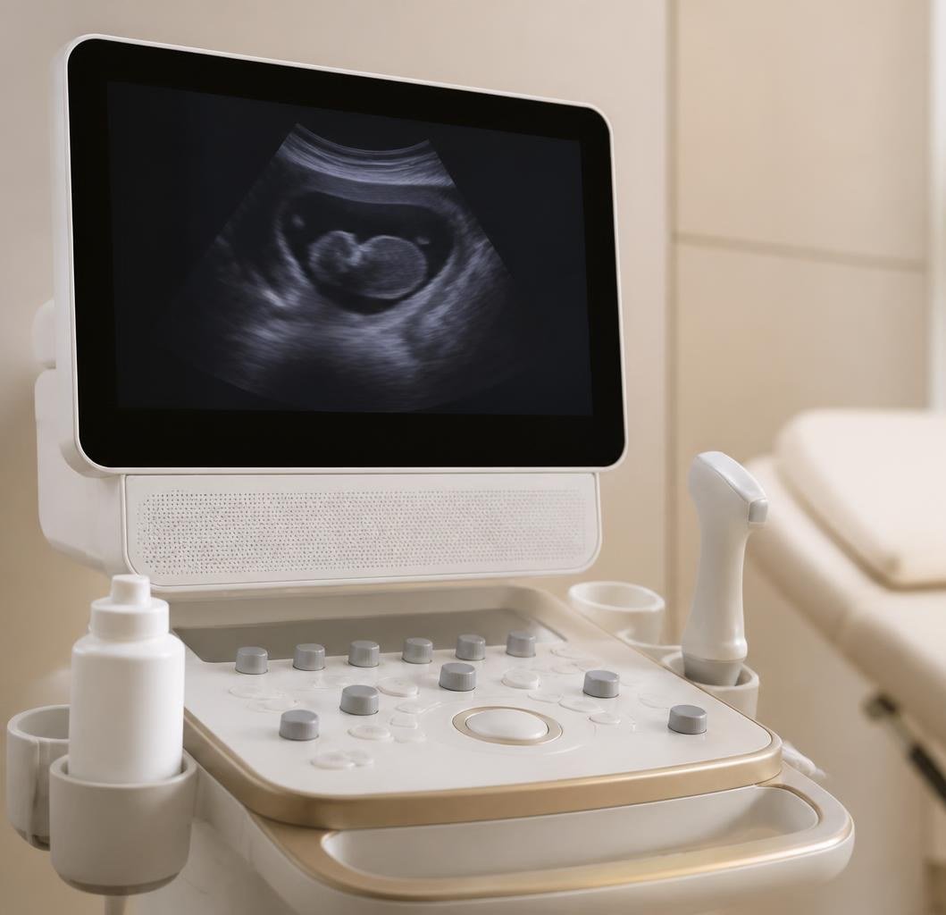
"And what is this one for?" It is the question I hear most when I hand over an ultrasound request. Many patients arrive knowing it is due, but without a clear idea of what is being looked for that day, or why that week and not another. Knowing changes the experience: you go from waiting for a nice picture to understanding what is being checked.
Each ultrasound has a specific time window. Outside it, the same scan stops answering the question it was meant to answer.
First trimester: weeks 11 to 14
This is the one that sets up everything else. It confirms the pregnancy is progressing and the heartbeat is present, tells you how many babies there are — worth knowing as early as possible — and pins down the due date to within a few days. It also measures nuchal translucency, a fold at the back of the baby’s neck whose thickness helps estimate the risk of certain chromosomal conditions.
- Confirms viability and the number of fetuses.
- Sets the due date with the greatest precision of the whole pregnancy.
- Measures nuchal translucency, valid only between weeks 11 and 14.
- Takes about 20 minutes.
Anatomy scan: weeks 20 to 22
If you could only have one, this would be it. The baby is reviewed organ by organ: the heart and its four chambers, brain, kidneys, spine, limbs, face, along with the placenta and the amount of amniotic fluid.
That is why it takes 30 to 45 minutes, considerably longer than a routine check, and why I sometimes ask you to walk around or come back another day: if the baby is lying in a position that hides a structure, it still needs to be seen. This is the point in pregnancy when malformations are most reliably detected, and that window does not come round again.
- Complete anatomical review, organ by organ.
- Assesses the placenta, cord and amniotic fluid.
- It is when the baby's sex can be seen most reliably.
- Takes 30 to 45 minutes and may require a second visit.
Third trimester: weeks 32 to 36
Here we are looking for something different. We check that the baby is growing at the expected rate, what position it is in — key to discussing the mode of delivery — where the placenta sits and whether amniotic fluid is adequate. This is the scan that tells us most about how the final weeks and the birth are likely to go.
What I am asked most
Do they harm the baby? No. Ultrasound is not radiation; it has been used in obstetrics for more than fifty years. When can we tell the sex? Sometimes it can be guessed from week 16, but it is reliably confirmed at the anatomy scan. What about 4D scans? They are lovely and many families enjoy them, but they are an extra: they do not replace any of the three and add no diagnostic information. Only three? In an uncomplicated pregnancy, yes. If something needs monitoring, we add as many as required.
A note about the dates
The weeks you have read here are not approximate: they are the windows in which each scan delivers what is expected of it. An anatomy scan at week 26 no longer sees what it would at week 21. That is why I insist so much on appointments, even when everything seems fine. If you are pregnant and have not started prenatal care yet, book your first visit: ideally between weeks 6 and 8.